Megan Brooks
July 21, 2016
A team of neuroscientists has charted the most complex and precise map of the human cerebral cortex to date, and it has revealed almost 100 new regions. These findings will provide a greater understanding, as well as advance the study, of brain disorders such as autism, schizophrenia, dementia, and epilepsy.
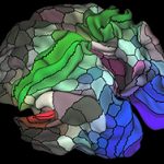
The map, dubbed Human Connectome Project Multi-Modal Parcellation version 1.0 (HCP-MMP1.0), divides each hemisphere of the brain into 180 distinct cortical regions; of these, 97 have never been described before.
The report was published online July 20 in Nature.
Imaging Data
The team, led by Matthew Glasser, PhD, and David Van Essen, PhD, of Washington University School of Medicine, in St Louis, Missouri, created the map using imaging data from 210 healthy young adults from the Human Connectome Project.
They combined several brain mapping techniques that have previously been used only separately, namely, task-based functional MRI (fMRI), which yields information on the functions of different regions; resting-state fMRI, which details neural connectivity within and between different regions; and relative density of the neuron-sheathing substance myelin, which provides data about cortical architecture.
A 180-area multimodal human cortical parcellation on the left and right hemisphere surfaces. (Credit: Matthew F. Glasser, David C. Van Essen)
“We used multiple measures of the organization of the cerebral cortex,” Dr Glasser told Medscape Medical News. “We looked at microstructural architecture, function, connectivity, and topographic maps. We focused on those locations where these maps changed in multiple independent measures, giving us more confidence in these areal boundaries,” he explained.
Next, the researchers trained a machine learning algorithm to recognize each cortical area on the basis of its multimodal “fingerprint” ― that is, its pattern of architecture, function, connectivity, and topographic maps, Dr Glasser said.
Despite individual variability, this algorithm successfully found these areas in an independent group of 210 study participants who were not part of the original parcellation or of the algorithm training.
First Application
Dr Glasser said the first application will be in brain imaging research. The map will help investigators know where they are in the cerebral cortex when they conduct a study.
“That will make it easier for them to compare with a colleague’s separate study to see if they found the same brain area activated, neighboring brain areas activated, or the activations were further away,” he said.
In the past, it was not always clear whether the results from two neuroimaging studies related to the same area or not. By using the new map and alignment algorithm, results of separate studies can be more accurately compared, he explained.
Individualized brain maps may also help neurosurgeons avoid damaging important parts of the brain, such as those that control movement or speech, Dr Glasser said.
“We were able to persuade Nature to put online almost 200 extra pages of detailed information on each of the 180 regions as well as all of the algorithms we used to align the brains and create the map,” Dr Van Essen commented in a news release. “We think it will serve the scientific community best if they can dive down and get these maps onto their computer screens and explore as they see fit.”
Springboard for Discovery
“These new insights and tools should help to explain how our cortex evolved and the roles of its specialized areas in health and disease, and could eventually hold promise for unprecedented precision in brain surgery and clinical workups,” Bruce Cuthbert, PhD, acting director of the National Institute of Mental Health, which cofunded the research as part of the Human Connectome Project, said in a statement.
“An authoritative map of the modules that make up the cerebral cortex of the human brain promises to act as a springboard for greater understanding of brain function and disease,” write B. T. Thomas Yeo, PhD, of the National University of Singapore, and Simon Eickhoff, MD, PhD, Heinrich-Heine University, Dusseldorf, Germany, in an accompanying editorial.
“This long-awaited advance provides a reference atlas that will allow those researching brain structure, function and connectivity to work within a common, systems-neuroscience framework.
“It is now up to researchers to use the anatomical framework provided, compare it with alternative approaches to mapping the human brain, and populate the defined areas with functional and disease related information,” they write. “By doing so, we can begin to integrate multimodal data to understand how individual differences in brain organization can explain differences in function, behaviour and disorder.”
Dr Yeo and Dr Eickhoff note, however, that the authors’ validation of this algorithm focused on only a small portion of the cortex, so further investigation is crucial.
“Nevertheless, their work represents a major step towards individual-specific ‘biomarkers’ of brain dysfunction, because individual-specific quantities of each area, such as grey-matter volume or connectional strength to other areas, can now be computed, and could be strongly predictive of individual differences in behaviour or disease,” they write.
Nature. Published online July 20, 2016. Abstract, Editorial